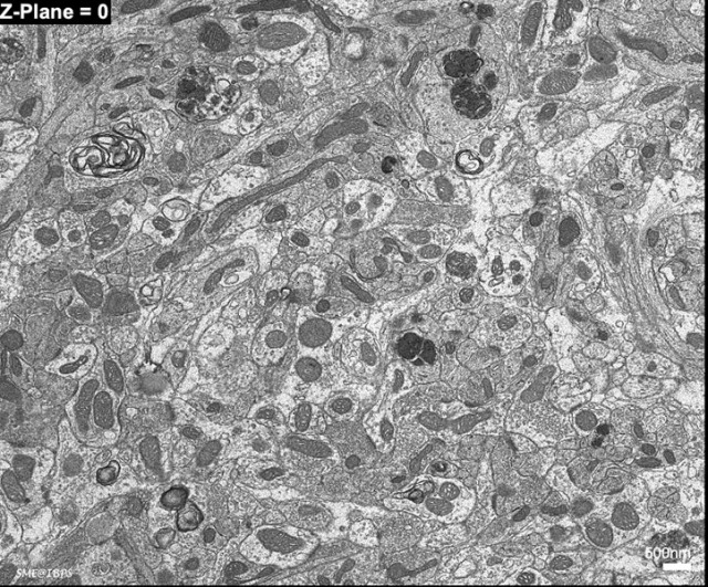
Electron Microscopy Facility

The Electron Microscopy Facility (EMF) of the Institut de Biologie Paris-Seine offers a set of skills and equipment in transmission and scanning electron microscopy (TEM, SEM) to the scientific community (IBPS, Sorbonne Université, public and private laboratories). A team of three engineers animate the EMS. They provide methodological supportuser trainingmaintenance of the equipment and scientific activities (seminars, teaching).

Our skills

EMF provides access to a wide range of equipment allowing the preparation and the observation in TEM and in SEM of a large variety of models, from bulk specimens or cell cultures to thin films or nanoparticles. Sample preparation dedicated to ultrastructural, immunolocalisations or 3D studies can be realised "conventionally", with chemical methods or using cryomethods. The facility has a long-lasting expertise in cryomethods, which associate a fast immobilisation of samples with an optimal preservation of their integrity (high-pressure freezing, freeze substitution, plunge-freezing, freeze-fracturing, freeze-etching, cryo-SEM).

Methodologies

  • Conventional sample preparation and observation in TEM: ultrastructural cytochemistry, immunocytochemistry, negative contrast
  • Freezing: high pressure freezing, plunge-freezing and freeze-substitution
  • Conventional sample preparation and observation in high-resolution SEM, cryo-SEM (freeze-fracturing and freeze-etching) and STEM-in-SEM
  • "Volumetric" imaging with Array Tomography
  • Correlative light electron microscopy (CLEM)

Services

Presentation of offers:

All projects developed within the platform may include a conception monitoring, follow-up, realisation and result analysis. An interview precedes each project to evaluate feasibility and to propose the most appropriate techniques.

Type of offers:

  • Sample preparation (room temperature and cryo) in TEM and SEM
  • Training in sample preparation and image acquisition
  • Engineer-assisted observation sessions
  • Observation sessions for autonomous users
  • Long-term collaborative projects

Material

Our park is equipped with sample preparation apparatus, a high-resolution Scanning Electron Microscope (SEM) to visualise the sample surface, and for array tomography. Furthermore it includes one Transmission Electron Microscope (TEM) to observe standard sections of samples, or to reach molecular level (80 - 200 kV).

  • Transmission Electron Microscope 200kV (2100HC, JEOL)
  • Field-Emission Scanning Electron Microscope (GeminiSEM 500, Zeiss) with cryo-stage (VCT100, Leica)
  • Vibratome (VT1000S, Leica)
  • Ultramicrotomes, cryo-ultramicrotome (UCT, UCS, Leica)
  • Plunge-freezing apparatus (CPC, Leica)
  • High pressure freezing (HPM100, Leica)
  • Freeze-substitution apparatus (AFS, Leica)
  • Critical point drying apparatus (CPD300, Leica)
  • High-resolution Coaters (ACE600, Leica)
  • Data treatment and tomographic reconstruction (Imod, TomoJ, Atlas5)

Methodological development

The service is involved in various technological developments to provide innovative methodological solutions, for example: optimal sample preservation (cryomethods), 3D reconstruction (array tomography) or screening for rare events detected with fluorescence (correlative microscopy).

Thereby, from January 2016, our platform is equipped with a high-resolution SEM (Zeiss GeminiSEM 500) coupled to cryopreparation (Leica VCT100) and automatic imaging of serial sections (Zeiss Atlas 5 AT). This will permit high-resolution viewing of various samples maintained in their native state, at the subcellular and molecular levels, and high contrast TEM-like imaging, with a very wide field of view and 3D visualization of large volumes in Array Tomography (3D reconstruction).

News of the platform

Collagen Like You’ve Never Seen it Before!

New biomaterials can be developped through the in vitro production of collagen membranes, according to a recent study led by Carolé Aimé. This publication was realized by the teams Matériaux et biologie and Reactive Materials for Electrochemical devices - part of the Condensed Matter Chemistry Laboratory in Paris (UMR 7574) which specializes in the engineering of biomaterials

The EMF contributed to the study’s multi-modal and multi-scale analyses by carrying out a rare and original imaging approach that uses "direct cryo-SEM". The sample was viewed on a nanoscopic scale in a fully hydrated and quasi-native state.

The sample only needs to be frozen in place (plunge-freezing) before observation at a very low temperature, a method which allows the preservation of both the organization and fine structure of the collagen fibrils. These can otherwise be greatly altered with conventional SEM sample preparation methods that require chemical preparation (fixation, dehydration, drying and surface coating).

The article, with acknowledements for EMF staff, can be downloaded here:

https://pubs.acs.org/doi/abs/10.1021/acsabm.0c00006

A High-resolution SEM to Explore Tissue Ultrastructure in Three Dimensions

SME_ArrayTomo_Mitoch

This example of nervous tissue (collaboration with the team Development and Plasticity of Neural Network, UMR 8246 Neurosciences) was used to explore parts of the olfactory neural layers of mice, applying the array tomography method.

Array tomography exploits the performance of the platform’s high-resolution Scanning Electron Microscope (SEM). The SEM - equipped with next-generation optics and detectors along with high-performing automation software (Atlas 5 Zeiss) - can produce "TEM type images" from conventionally prepared biological samples (fixation, inclusion, ultramicrotomy and contrast).
The images obtained have a nanometric lateral resolution (here 2.5 nm / pixel) and an axial resolution resulting from the thickness of the different ultra-fine sections recovered (here 80 nm).

Array tomography coupled with our high-performance SEM makes it possible to obtain:

  • observations of millimetric fields of view, using a mosaic approach with the acquisitions of contiguous regions
  • sample volumes covering a depth of several micrometers, acquiring the same regions of interest repeated over tens / hundreds of successive sections